![[Achnanthes], X640.](/assets/Tools-And-Resources/Identification/algae/achnanthes__ResizedImageWzQwNSwyODld.jpg "[Achnanthes], X640.")
[Achnanthes], X640.
Diatoms are characterised by a unique feature: a cell wall composed of silica, which fits together in two halves like a box. The lid and base of the box are known as valves, which are connected by a girdle, and the whole structure is known as a frustule. This has the important consequence that a cell can appear quite different if viewed from the side ("girdle view") than if it is viewed from above ("valve view"). However, once this cell architecture is understood, a great world of diatom diversity becomes accessible to the investigator.
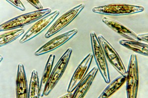
Frustules occur in two basic forms, cylindrical ("centric", circular in valve view) and elongated ("pennate", roughly boat-shaped in valve view, although there are many variations on the theme). The centric form is the oldest; all modern diatoms are ultimately descended from ancient centric diatoms. An extremely common centric diatom in freshwaters is Melosira, which forms long filaments of cells joined at the valve faces. An example of a pennate diatom is an unfortunate recent arrival in New Zealand, Didymosphenia, known also as "Didymo" or "rock snot".
Diatom frustules are sculpted in various ways by openings that pass through the silica. Striae, which are lines of pores, are commonly visible under the light microscope. More useful to identify the taxa in this guide is the raphe. This is a feature of some pennate diatoms. It is a slit-like structure extending lengthwise along the cell and that extrudes mucilage, allowing cells to glide along surfaces such as a microscope slide. Some pennate diatoms have raphes on both valve faces (e.g., Navicula), some on one (e.g., Planothidium), and some on neither (e.g., Ulnaria). In some genera the raphes are positioned in the centre of the valve face (e.g., Navicula), in others it is positioned to the side (e.g., Nitzschia), and in still others it traverses from the valve face onto the side of the valve (e.g., Eunotia). Therefore, an appreciation of this structure is very helpful for accurate identification.
Diatoms contain golden-brown chloroplasts, and contain chlorophylls a and c (never chlorophyll b). With practice, they can often be recognised in the field by their colour. Serious diatomists clean the cells with acid and peroxide so that only the frustules remain, aiding identification. However, many useful features can still be recognised in living material, and images in this guide often compare living and cleaned cells for comparison (see here for an example).
![[Achnanthes], Lake Waihola, X640. Photo: Otago Regional Council & Manaaki Whenua](/assets/Tools-And-Resources/Identification/algae/achnanthes__FillMaxWzMwMCwyMDBd.jpg)
![[Cerataulina], lower Taieri River, X160. Photo: Otago Regional Council & Manaaki Whenua](/assets/Tools-And-Resources/Identification/algae/cerataulina160__FillMaxWzMwMCwyMDBd.jpg)
![[Cyclotella], X1000. Photo: Phil Novis, Manaaki Whenua](/assets/Tools-And-Resources/Identification/algae/cyclotella_meneghiniana_phil1000__FillMaxWzMwMCwyMDBd.jpg)
![[Cymbella], X400. Photo: Manaaki Whenua](/assets/Tools-And-Resources/Identification/algae/cymbella400__FillMaxWzMwMCwyMDBd.jpg)
![[Diatoma], X1000. Photo: Phil Novis, Manaaki Whenua](/assets/Tools-And-Resources/Identification/algae/diatoma_phil1000__FillMaxWzMwMCwyMDBd.jpg)
![[Didymosphenia] on mucilaginous stalks, X200. Photo: Manaaki Whenua](/assets/Tools-And-Resources/Identification/algae/didymo200__FillMaxWzMwMCwyMDBd.jpg)
![[Eunotia sepentina], north Taranaki, X400. Photo: Taranaki Regional Council & Manaaki Whenua](/assets/Tools-And-Resources/Identification/algae/eunotia2__FillMaxWzMwMCwyMDBd.jpg)
![[Melosira], X1000. Photo: Otago Regional Council & Manaaki Whenua](/assets/Tools-And-Resources/Identification/algae/melosira3__FillMaxWzMwMCwyMDBd.jpg)
![[Nitzschia], Kaikorai tributary, X400. Photo: Otago Regional Council & Manaaki Whanua](/assets/Tools-And-Resources/Identification/algae/nitzschia2__FillMaxWzMwMCwyMDBd.jpg)
![[Planothidium], X1000. Photo: Phil Novis, Manaaki Whenua](/assets/Tools-And-Resources/Identification/algae/planothidium_phil1000__FillMaxWzMwMCwyMDBd.jpg)
![[Ulnaria], X320. Photo: Otago Regional Council & Manaaki Whenua](/assets/Tools-And-Resources/Identification/algae/ulnaria320b__FillMaxWzMwMCwyMDBd.jpg)